The innovation: Safely sampling the thalamus during SEEG analysis
Researchers at Children’s Medical Center Dallas, part of Children’s HealthSM, have completed the first study in pediatric patients demonstrating the safety of sampling thalamic nuclei during stereo-electroencephalography (SEEG).Direct sampling enables neurologists to verify which nuclei are involved in a child’s seizures and plan more accurate treatment.
The study, published in the Journal of Neurosurgery: Pediatrics, included 10 patients with drug-resistant epilepsy (DRE) who underwent SEEG to map their seizure onset zones. Patients with multiple onset zones are ineligible for surgery and often depend on neuromodulation for treatment.
While neurologists commonly target thalamic nuclei for neuromodulation, they rarely sample these nuclei in SEEG analysis beforehand because of perceived risks to the patient. As a result, treatment may focus on the wrong nuclei and be ineffective.
“Neurologists no longer have to guess about thalamic involvement prior to treatment. We can sample it safely and use that data to choose neuromodulation strategies that may be more likely to reduce a child’s seizures,” says Angela Price, M.D., Pediatric Neurosurgeon at Children's Health and Associate Professor at UT Southwestern, who led the research.
The big picture: Neuromodulation depends on accurate assessment of onset zones
About 25% of children with epilepsy have DRE and must be considered for either surgery or neuromodulation. Among neuromodulation methods, Dr. Price favors responsive neurostimulation (RNS) because it’s a closed-loop system: An array of electrodes both monitor brain activity and deliver electrical impulses to interrupt seizures as they occur. Open-loop systems like deep-brain stimulation (DBS) and vagal nerve stimulators don’t monitor, they deliver impulses on a steady cadence, like a pacemaker.
“But as great as RNS is, about 1 in 4 people with DRE don’t benefit from it,” Dr. Price adds. One reason could be the placement of the electrodes: To interrupt seizure activity, electrodes must target parts of the brain where seizures originate. The thalamus may be one of those places that is involved in the seizure network– but it is also prime cerebral real estate, involved in every major function of the brain. Many neurosurgeons worry that an invasive procedure like SEEG is too dangerous to make sampling the thalamus worthwhile.
So rather than test the thalamus firsthand, they assume thalamic involvement in seizures based on broader regions of the brain that show activity: If the frontal cortex shows activity, for example, they target the anterior nucleus of the thalamus (ANT) for treatment. “To help more children with DRE, we need to move beyond assumptions. We need to know exactly where their seizures are coming from, or the networks that are active” Dr. Price says.
Key details: Sampling the thalamus is safe and data is often surprising
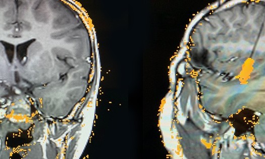
For the patients in their study, Dr. Price and team sampled a one or more thalamic nucleus: the left ANT, the right ANT, the left centromedian nucleus of the thalamus (CMT), or the right CMT. Seven patients had a single nuclei sampled, two patients had two nuclei sampled and one patient had three. Patients were monitored between four and nine days, depending on the frequency of their seizures.
The safety results were compelling:
No hemorrhages in the thalamus or along the thalamic electrode.
No infections.
No strokes.
Seven patients proved to have multiple seizure onset zones requiring neuromodulation – but the team’s assumptions about the involvement of thalamic nuclei were wrong almost half the time: In three cases, the thalamic nuclei selected for sampling played no role in the child’s seizure network.
“Without sampling the thalamus during SEEG, we would have targeted those nuclei for RNS treatment and the child may have received no benefit,” Dr. Price explains.
How to perform SEEG safely: Robotics and proper MRIs
The seven patients with multifocal seizures have proceeded to treatment with RNS. The team selected thalamic targets based on individual SEEG findings: The team will follow the patients and evaluate at two years to see if this approach to selecting thalamic targets is effective at reducing seizures.
In the meantime, Children’s Health now samples the thalamus for all patients undergoing SEEG in which neuromodulation may be indicated following SEEG (patients that are unlikely to be a candidate for resective brain surgery). Very few pediatric centers follow this practice, but Dr. Price encourages all centers with SEEG capability to do so.
Here’s how:
Prior to SEEG, all patients should undergo a complete phase 1 evaluation and be discussed at a comprehensive epilepsy conference. If a candidate for SEEG, and likely to find a resective lesion, sampling of the thalamic nuclei should be considered.
Use appropriate MRI to plan trajectories and enable direct targeting:
T1 with contrast to reveal blood vessels.
MRI FGATIR and MP2RAGE to visualize the thalamic nuclei.
During SEEG, Robotic Surgical Assistant (ROSA) laptop can assist to guide electrode placement.
To learn even more during SEEG, Dr. Price recommends using the thalamic electrodes to stimulate the implanted nuclei and see whether it produces a seizure. Dr. Price and team tried this with two patients, and for one of them a seizure did result. “Then you know you found a hot spot. And your patient hopefully will be a high RNS responder, we will see if this is supported with further studies,” says Dr. Price.
Learn more about innovations in pediatric epilepsy case at Children's Health.